INTRODUCTION
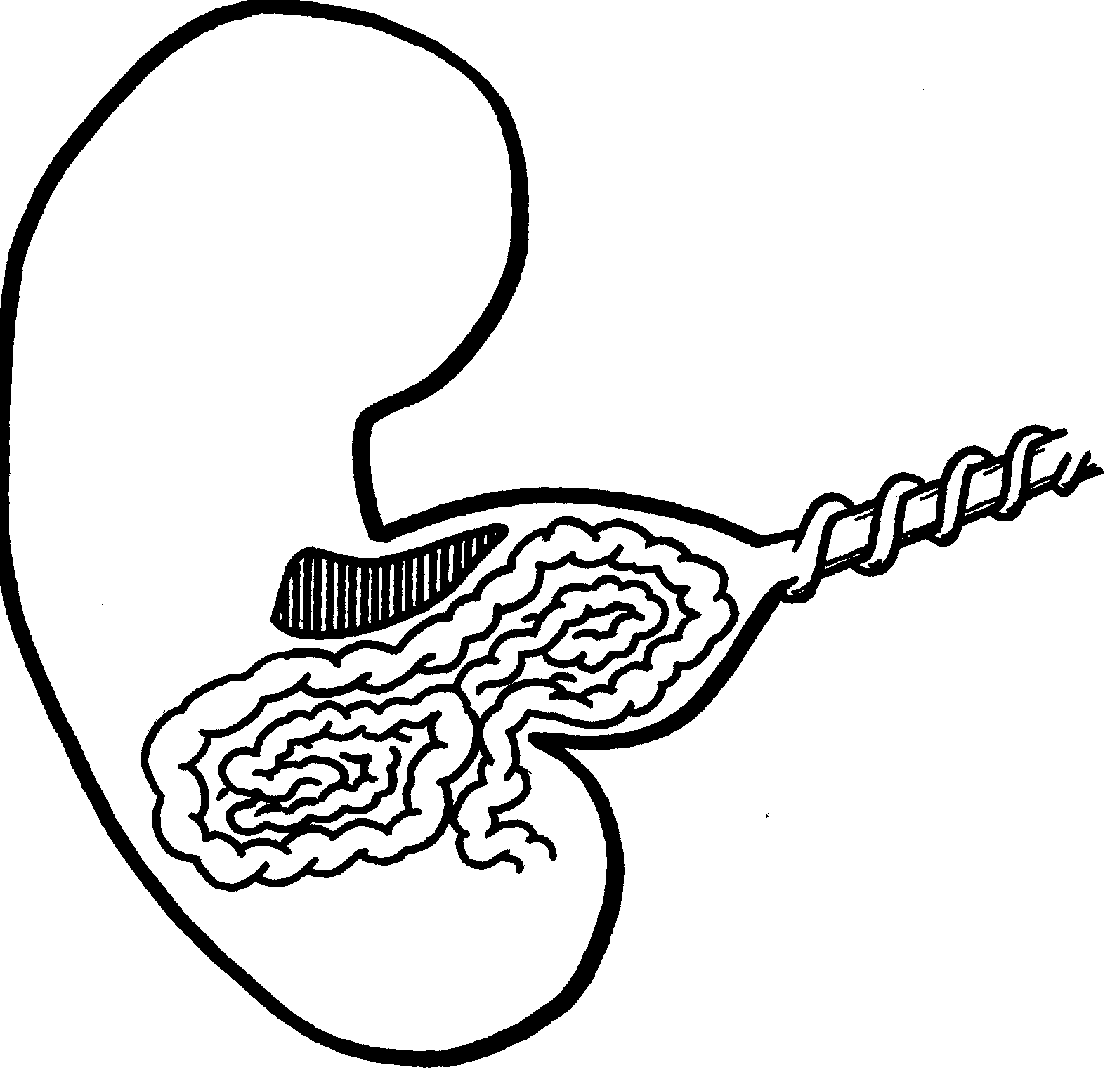
| In an exomphalos some of the abdominal
contents are found outside the abdomen in a thin
clear sac to which the baby's umbilical cord is
attached. The sac consists of amnion and parietal
peritoneum with some mesenchyme between. A small
exomphalos may contain only a Meckel's diverticulum
whilst a large defect may contain the stomach,
liver and bladder. Non-rotation of the intestines
is commonly seen. |
 |
Exomphalos is frequently associated with major abnormalities
of other systems suggesting that this anomaly is not
a simple failure of umbilical ring closure. The primary
defect is likely to occur in early development. Most
cases of exomphalos are sporadic, however the condition
is often associated with aneuploidy and familial occurrence
has been described. Exomphalos associated with macroglossia
and macrosomia is known as Beckwith-Wiedemann syndrome.
Go to the top of this
page
ANTENATAL
Most cases are identified by routine fetal anomaly
scans and may be associated with elevated maternal
serum alphafetoprotein (AFP) levels. On ultrasound
the normal cord insertion is not seen but is replaced
by a mass representing the bowel or liver herniating
into the base of the umbilical cord. The mass encapsulated
by a membrane is seen to be attached to the cord. Careful
ultrasound examination will usually identify coexistent
structural anomalies (especially cardiac anomalies)
which exist in 70-85 % of fetuses.
Karyotyping should always be offered as at least
35% of fetuses have a chromosome abnormality. The presence
of other abnormalities including aneuploidy will largely
determine the prognosis. Frequently, a poor prognosis
is predicted and termination of pregnancy requires
discussion. Serial ultrasound measurements should be
performed noting fetal growth if the pregnancy is continued.
Go to the top of this
page
POSTNATAL
In cases with a normal karyotype and no major associated
anomalies a vaginal delivery may be appropriate. Large
lesions, especially containing liver are delivered
by caesarean section. Detailed prenatal ultrasound
may not identify all potential problems making a careful
neonatal assessment vital in these cases. In choosing
the place of delivery, consideration should be given
to the availability of fetal medicine, neonatal and
specialist surgical expertise.
With a small or medium sized exomphalos one operation
to close the abdomen is all that is usually required.
Sometimes the skin but not the muscles can be closed
over an exomphalos sac and later operations are needed
to close the muscles. If the sac is large, a silastic
pouch can be used to allow progressive reduction of
the bowel over a number of days before closure. Very
large sacs can also be managed without operation. The
sac can be painted with agents that lead to a scar
forming, over which skin slowly grows. If skin alone
has been used to cover the defect a ventral hernia
results, and subsequent procedures are required to
prepare this. Survival depends on the other defects
and varies from 30-70%.
Go to the top of this
page
WEST MIDLANDS
DATA

Go to the top of this
page
|

