INTRODUCTION
Trisomy 13 is the
most severe viable trisomy caused by an additional
copy of chromosome 13. Most pregnancies will result
in miscarriage but some survive the first few weeks
of life. As with other trisomies such as Down's
syndrome there is a maternal age effect with an
increased incidence in older mothers.
Additional structural anomalies
are common, particularly facial anomalies (midline
clefts, hypotelorism, microphthalmia, and anophthalmia)
arising from structural anomalies of the brain,
frequently microcephaly and holoprosencephaly.
Other associated anomalies include cardiac, renal,
and intestinal (diaphragmatic hernia) anomalies.
Characteristic features include low set ears, post-axial
polydactyly, flexion contractures, rocker bottom
feet, scalp defects, and haemangiomas.
Top of Page
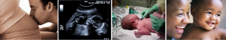
ANTENATAL
As with trisomy 18 many fetuses
affected by trisomy 13 will show early intrauterine
growth failure, and will measure smaller than expected
at the first scan. Major structural anomalies are
occasionally identified in the late first or early
second trimester, but the most common initial presentation
is for the small measurement to be interpreted
as incorrect dating of the pregnancy. Subsequent
scans again indicating poor fetal growth indicate
a more serious underlying problem.
Other associated anomalies will be visible on ultrasound at 20 weeks including
facial clefts, holoprosencephaly, cardiac defects, diaphragmatic hernia, and
hexadactyly. The discovery of a structural anomaly in association with growth
failure increases the chances of chromosomal anomalies, and women should be
offered amniocentesis or other invasive testing to establish the fetal karyotype.
Following a confirmed prenatal diagnosis, the decision to be made is either
to terminate the pregnancy, or to manage it conservatively.
If a fetus with significant structural
anomalies dies in-utero, the medical staff should
be suspicious of a chromosomal anomaly. In women
who have elected not to have invasive testing the
question should be asked again, as there is a much
higher success rate with amniocentesis, than waiting
for post delivery cultures, which often fail due
to bacterial over-growth. In contrast, fetal cells
can usually be cultured, even several days after
intra-uterine death.
Couples who have suffered the loss
of a child with Patau's syndrome should be offered
invasive testing in any future pregnancy. The recurrence
risk is small, and is not precisely known and many
couples will decline in the knowledge that ultrasound
can give a degree of reassurance for this condition,
and that invasive testing carries a small risk
of pregnancy loss.
Top of Page
POSTNATAL
There is a high rate of attrition
of affected pregnancies and many abort spontaneously.
Trisomy 13 is reported to be as common in spontaneous
abortions as trisomy 18. Most affected live born
infants die within the first weeks of life. Some
cases with mosaic trisomies have survived for several
years, all affected by severe mental retardation.
Top of Page
WEST MIDLANDS
DATA
Information to follow