Definition
An encapsulated fluid
filled space within the choroid plexus of the developing
fetal cerebral ventricle.
Explanation
The choroid plexus is
a bundle of blood vessels with can be clearly seen
within the cerebral ventricles. The blood vessels
appear echogenic, but cysts can form within these
developing blood vessels. Choroid plexus cysts (CPCs)
can be unilateral or bilateral, but are generally
best seen on ultrasound in the far field – the
side of the head furthest away from the scan probe.
Technique for
measurement/assessment
A CPC is seen in the standard
view for the cerebral ventricles. A transverse section
including both choroids should be obtained, and the
image showing the CPC most clearly should be measured
and assessed. The CPC should be imaged in 3 planes
and seen to lie within the developing choroid. The
largest diameter of the CPC should be measured. It
is common for CPCs to be bilateral, but the cysts
lying in the near field may be more difficult to
image. There should be no change in the appearance
of the lateral ventricles, or other structures in
the fetal brain. The ultrasound assessment should
be completed, looking in particular for the other
soft markers: nuchal pad, echogenic bowel, echogenic
foci, short femur length and renal pyelectasis.
Implications of
the finding
Choroid plexus cysts are
associated more with trisomy 18 (Edward’s syndrome)
than trisomy 21 ( 1)(Down’s
syndrome). CPCs are most common at 16 weeks gestation,
when about 2% of fetuses will demonstrate this finding,
and almost always disappear by 26 weeks (2).
Trisomy 18 has a high chance (80%) of demonstrating
other ultrasound features. Hydrocephalus, holoprosencephaly,
midline facial cleft, cardiac anomalies, diaphragmatic
hernia, exomphalos, clenched hands with overlapping
fingers, talipes and early onset growth failure should
be sought (3).
The fetal hands in particular have been identified
as an important feature to distinguish a case of
trisomy 18 (4) and
therefore normal, open hands should be seen in conjunction
with normal anatomy and growth for a diagnosis of
an isolated CPC to be made. 1)(Down’s
syndrome). CPCs are most common at 16 weeks gestation,
when about 2% of fetuses will demonstrate this finding,
and almost always disappear by 26 weeks (2).
Trisomy 18 has a high chance (80%) of demonstrating
other ultrasound features. Hydrocephalus, holoprosencephaly,
midline facial cleft, cardiac anomalies, diaphragmatic
hernia, exomphalos, clenched hands with overlapping
fingers, talipes and early onset growth failure should
be sought (3).
The fetal hands in particular have been identified
as an important feature to distinguish a case of
trisomy 18 (4) and
therefore normal, open hands should be seen in conjunction
with normal anatomy and growth for a diagnosis of
an isolated CPC to be made.
Trisomy 18 is associated
with advanced maternal age and a low HCG on serum
screening (in contrast to trisomy 21 where the serum
HCG is high). Therefore women of >35 years of
age with an isolated CPC should be considered high
risk for trisomy 18 even if serum screening gives
a low risk for trisomy 21 (5).
If there are no other
problems or risk factors there is no need for further
action. The cysts usually disappear, and although
possibly reassuring in terms of the growth of the
fetus, there is no evidence that repeating ultrasound
scans later in the pregnancy affects the outcome.
If a further scan is to be undertaken it is logical
to do this in the third trimester (28 to 34 weeks)
to identify growth failure which is commonly associated
with trisomy 18. There seems little to be gained
by repeating scans earlier than this unless the anatomical
structural survey, or markers checklist remains incomplete.
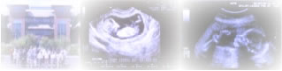
Image 6- Choroid
plexus cyst

References
1. Fitzsimmons J, Wilson
D, Pascoe-Mason J, Shaw CM, Cyr DR, Mack LA. Choroid
plexus cysts in fetuses with trisomy 18. Obstetrics
and Gynecology. 1989; 73: 257-260, Abstract
2. Morocos CL, Platt
LD, Carlson DE, Gregory KD, Greene NH, Korst LM.
The isolated choroids plexus cyst. Obstet Gynecol
1998; 92: 232-236, Abstract
3. Gupta JK, Khan KS,
Thornton JG, Lilford RJ. Management of fetal choroids
plexus cysts. British Journal of Obstetric and Gynecology.1997;
104:881-886, Abstract
4. Sahinoglu Z, Uludogan M, Sayar C, Turkover B,
Toksoy G. Second trimester choroid plexus cysts & trisomy
18. Int J Gynaecol Obstet 2004 Apr; 85(1): 24-9, Abstract
5. Chitty LS, Chudleigh P, Wright E, Campbell S,
Pembrey M. The significance of choroid plexus cysts
in an unselected population: results of a multicenter
study. Ultrasound Obstet Gynecol 1998 Dec; 12(6):
391-7, Abstract
|