Definition
Thickening ³
6 mm in the posterior aspect of the neck.
Explanation
This should not be confused with nuchal
translucency which is a distinct measurement taken
at 10-14 weeks. Neither should it be confused with
a cystic hygroma, which is a septated, fluid filled
structure around the fetal neck.
A thickened nuchal pad may be an early
sign of hydrops but is usually an incidental finding.
Standard image for identification/exclusion
Transverse view of the cranium across
the thalami, angled slightly posteriorly to include
the cerebellum and occipital bone.
Technique for measurement/assessment
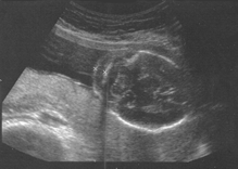
The nuchal pad should be measured by
placing one cursor at the outer edge of the occipital
bone and the other at the skin surface.
The ultrasound assessment should be
completed, looking in particular for the other soft
markers: echogenic bowel, echogenic foci, short femur
length, renal pyelectasis and choroid plexus cysts.
Signs of hydrops should also be sought.
Implications of a positive
finding in isolation
The recent meta-analysis by Smith-Bindman
et al suggests that the positive likelihood ratio
for nuchal pad is 17 times the background risk for
trisomy 21. This will result in the vast majority
of people with this finding being considered high
risk and justifies a discussion of karyotyping.
The consensus view at this time is for karyotyping to be discussed and offered,
but we are aware of the deficiencies in the current evidence. Audit findings from the West Midlands data during 2000-05 support offering karyotyping for isolated nuchal pad.
Image 1 - Nuchal Pad
References
1. Nyberg DA, Luthy DA, Resta RG, Nyberg
BC and Williams MA. Age-adjusted ultrasound risk
assessment for fetal Down’s syndrome during
the second trimester: description of the method and
analysis of 142 cases. Ultrasound Obstet Gynecol
1998; 12 (1): 8-15, Abstract
|