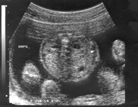
Definition
Dilatation of the fetal
renal pelvis in AP diameter 5-10 mm, with no calyceal
involvement.
Explanation
The renal pelvis connects
the renal calyces and ureter. By 16 weeks the amniotic
fluid consists of fetal urine reflecting a functional
renal system. Dilatation may reflect a downstream
obstruction or intrinsic laxity in the collecting
system and is often a variant of normal.
Standard image
for identification/exclusion
A transverse section
of the fetal abdomen should be obtained and one kidney
identified in cross section. The transducer should
be rotated through 90° to obtain a longitudinal
section and the appearance of the renal pelvis should
be noted. Dilated renal calyces should be looked
for; if present these are confluent with the renal
pelvis. The finding of dilated calyces, renal pelvis
dilatation exceeding 10 mm or ureteric dilatation
are usually abnormal and warrant a detailed ultrasound
scan with or without tertiary referral.
Technique for
measurement/assessment
The maximum AP diameter
of the kidney and renal pelvis should be made in
the transverse plane. The fetal bladder and liquor
volume should also be examined.
The ultrasound assessment should be completed, looking in particular for the
other soft markers: nuchal pad, echogenic bowel, echogenic foci, short femur
length and choroid plexus cysts.
Implication of
a positive finding in isolation
Fetal pyelectasis may
indicate urinary tract obstruction, vesico-ureteric
reflux ( 1)or
normal variant. It may be an early sign of fetal
hydronephrosis or be a marker for abnormalities such
as renal duplication or reflux, which cannot be demonstrated
by ultrasound antenatally. However, fetal pelvic
dilatation is more common in the presence of maternal
pelvic dilatation (2)and
may reflect a common locally acting factor rather
than a distinct pathology. 1)or
normal variant. It may be an early sign of fetal
hydronephrosis or be a marker for abnormalities such
as renal duplication or reflux, which cannot be demonstrated
by ultrasound antenatally. However, fetal pelvic
dilatation is more common in the presence of maternal
pelvic dilatation (2)and
may reflect a common locally acting factor rather
than a distinct pathology.
There is a weak association
with trisomy 21 in conjunction with other markers,
but in isolation no further action is required.
In the absence of associated
abnormalities, a third trimester ultrasound should
be performed followed by postnatal ultrasound at
least 3-4 days after birth, repeated at between one
to three months of age (3)and
paediatric follow up.
Image 5 - Fetal
pyelectasis
References
1. Stamillo DM, Morgan
MA. Diagnosis of fetal renal anomalies. Obstet Gyn
Clin N Am 1998; 25:527-52, Abstract
2. Graif M, Kessler A,
Hart S, Daitzchman M, Mashiach S, Boichis H, Itzchak
Y. Renal pyelectasis in pregnancy: correlative evaluation
of fetal and maternal collecting systems. Am J Obstet
Gynecol 1992; 167: 1304-6, Abstract
3. Langer B. Fetal pyelectasis.
Ultrasound Obstet Gynecol 2000; 16:1-5, Abstract
|