Definition
An area of the fetal bowel with homogenous
echogenicity equal to that of the surrounding bone.
Explanation
The reason for this appearance has
yet to be elucidated and is likely to be multi factorial
to reflect the different associations. It has been
attributed to altered meconium composition ( 1),
bowel wall ischaemia (2)
and swallowed blood (3).
The incidence is 0.2-0.6%.
1),
bowel wall ischaemia (2)
and swallowed blood (3).
The incidence is 0.2-0.6%.
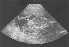
Standard image for identification/exclusion
The echogenicity should be at least
equal or greater than that of surrounding bone. There
should be no shadowing or enhancement to confuse
the appearances. The bowel must appear echogenic
in transverse and longitudinal sections of the abdomen.
Technique for measurement/assessment
Echogenic bowel with other signs
of placental failure will require further growth/liquor/Doppler
assessment.
Karyotyping should be considered by the Consultant reviewing the case.
Cystic fibrosis (CF) should be considered and parental blood taken for carrier status to identify couples at risk. If amniocentesis is taken for chromosome analysis CF studies can be requested. Any DNA analysis will only detect known mutations, currently 85% of clinical cases. All babies born in the UK are screened for CF after birth.
Blood should be taken for TORCH screen and parvovirus serology.
Image 2 - Echogenic Bowel
References
1. Nyberg DA, Dubinsky T, Resta RG,
Mahony BS, Hickok DE, Luthy DA. Echogenic fetal
bowel during the second trimester: clinical importance.
Radiology 1993; 188: 527-31, Abstract
2. Ewer AK, McHugo JM, Chapman S,
Newell SJ. Fetal echogenic gut: a marker of intrauterine
gut ischaemia? Arch Dis Child 1993; 69: 510-3, Abstract
3. Sepulveda W, Hollingsworth J,
Bower S, Vaughan JI, Fisk NM. Fetal hyperechogenic
bowel following intra-amniotic bleeding. Obstet
Gynecol 1994; 83:947-50, Abstract
4. Sepulveda W, Sebire NJ, Fetal
echogenic bowel: a complex scenario. Ultrasound
Obstet Gynecol. 2000; 16: 510-514, Abstract
5.
Sepulveda W, Nicholaides P, Mai AM, Hassan J,
Fisk NM. Is isolated second-trimester
hyperechogenic bowel a predictor of suboptimal
fetal growth? Ultrasound in Obstetrics & Gynecology.
1996;7(2):104-7, Abstract
6. Nyberg DA, Souter VL, El-Bastawissi A, Young
S, Luthhardt F, Luthy DA. Isolated sonographic markers
for detection of fetal Down syndrome in the second
trimester of pregnancy. J Ultrasound Med 2001 Oct;
20(10): 1053-63, Abstract
7. Al-Kouatly HB, Chasen ST,
Streltzoff J, Chervenak
FA. The clinical significance of fetal echogenic
bowel. Am J Obstet Gynecol 2001 Nov; 185(5): 1035-8,
Abstract