Definition
A small area within the fetal heart
with echogenicity equal to or greater than the surrounding
bone.
Explanation
There is histological evidence that
these areas represent calcification often within
the papillary muscle( 1),
the underlying reason for their presence is unknown
(2). 1),
the underlying reason for their presence is unknown
(2).
Standard image for identification/exclusion
In a standard four chamber view of
the heart, echogenic foci are seen in close proximity
to papillary muscles and should move in synchrony
with the valve leaflets.
They should be verified in a long axis
view. They are most common in the left ventricle
but can be found in the right or bilaterally. They
are discrete lesions and should not be confused with
diffuse areas of echogenicity, which are an indication
of a more general problem in the myocardium (3).
Technique for measurement/assessment
The number and location of the foci
should be assessed.
The ultrasound assessment should be
completed, looking in particular for the other soft
markers: nuchal pad, echogenic bowel, short femur,
renal pyelectasis and choroid plexus cysts.
Implications for a positive
finding in isolation
There is no association with cardiac
defects and referral for echocardiography is not
required in the absence of other indications. There
is no evidence that they affect cardiac function.
A prevalence of 3-5% has been described in the low
risk population (4)
and most will disappear prior to delivery or in the
early neonatal period.
There is a weak association with trisomy
21 in conjunction with other markers, but in isolation
no further action is required.
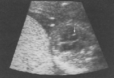
Image 3 - Echogenic foci in
the fetal heart
References
1. Robert DJ, Genest D. Cardiac histologic
pathology characteristic of trisomies 13 and 21.
Hum Pathol 1992; 23: 1130-40, Abstract
2. Pathologic correlation of sonographic
echogenic foci in the fetal heart. Prenat Diagn 2000;
20: 287-292, Abstract
3. Bronshtein M, Jakovi P Ofrir C.
Multiple fetal intracardiac echogenic foci:not always
a benign sonographic finding. Prenat Diagn 1996; 16:131-5, Abstract
4. Achiron R, Lipitz S, Gabbay U, Yagel
S. Prenatal ultrasonographic diagnosis of fetal heart
echogenic foci: no correlation with Down syndrome.
Obstet Gynecol 1997; 89:945-8 , Abstract
|