Definition
Femur length less than the third centile.
Explanation
The femur is the longest bone and
is measured as part of the routine mid pregnancy
anomaly scan. The distribution of limb length in
a population of fetuses will vary, but some will
be considered small. The definition used above allows
for local criteria and individual customisation.
Standard image for identification/exclusion
and technique for measurement/assessment
The femur should be measured with
the femur perpendicular to the ultrasound beam and
with the epiphyseal cartilages visible on the image.
The epiphyseal cartilages should not be included
in the measurement. Vertical measurement will foreshorten
the result due to increased velocity of ultrasound
in dense structures.
The ultrasound assessment should be
completed, looking in particular for the other soft
markers: nuchal pad, echogenic bowel, echogenic foci,
renal pyelectasis and choroid plexus cysts.
Implication of a positive finding
in isolation
This may be indicative of a general
skeletal malformation ( 1)
or growth problem. A full long-bone survey and examination
of the fetal chest should be performed. The foot
length is useful in interpretation of long bone measurement
(2).
Lethal malformations may be associated with a small
chest, which causes pulmonary hypoplasia. 1)
or growth problem. A full long-bone survey and examination
of the fetal chest should be performed. The foot
length is useful in interpretation of long bone measurement
(2).
Lethal malformations may be associated with a small
chest, which causes pulmonary hypoplasia.
Further management of an isolated
short femur may include growth assessment with ultrasound
later in pregnancy.
There is a weak association with trisomy
21 in conjunction with other markers, but in isolation
no further action is required.
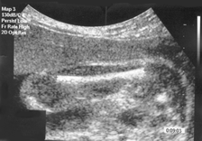
Image 4 - Short Femur length
References
1. Gonclaves L, Jeanty P. Fetal biometry
of skeletal dysplasias: a multicentre study. Journal
of Ultrasound in Medicine. 1994;13(12): 977-85, Abstract
2. Grandjean H, Sarramon MF. Femur/foot
length ratio for detection of Down syndrome: results
of a multicentre prospective study. The Association
Francaise pour le Depistage et la Prevention des
Handicaps de l’Enfant Study Group. American
Journal of Obstetric & Gynecology. 1995; 173(1):
16-9, Abstract
|